
Nati-1995/Brain-Tumor-MRI-Classification
Nati-1995/Brain-Tumor-MRI-Classification has 47 GitHub stars and 2 forks. GitHot measured 0 stars in 24 hours and +1 in 7 days. Last checked 2026-11-21 07:16 UTC. Last push 2026-08-16.
Progression mesurée des étoiles
- in 24h
- 0
- in 7 days
- +1
- in 15 days
- Historique en cours
- in 1 month
- Historique en cours
Variations nettes sur des périodes qui se chevauchent, pas des totaux quotidiens. Les mesures absentes restent vides. Les étoiles indiquent l’intérêt, pas la qualité.
À propos
Deep learning for brain tumor MRI: detection (99.4%) and 4-class typing (94.75%) via EfficientNet transfer learning + Grad-CAM.
Apprentissage automatiqueREADME.md
OriginalBrain Tumor MRI Classification
Deep-learning classification of brain MRI scans using transfer learning, with Grad-CAM interpretability to verify the model attends to actual tumor regions. Two tasks are covered: binary (tumor vs. no-tumor) and 4-class tumor typing (glioma, meningioma, pituitary, no-tumor).
Note: This is a research and educational project. It is not a medical device and must not be used for real diagnosis.
Demo
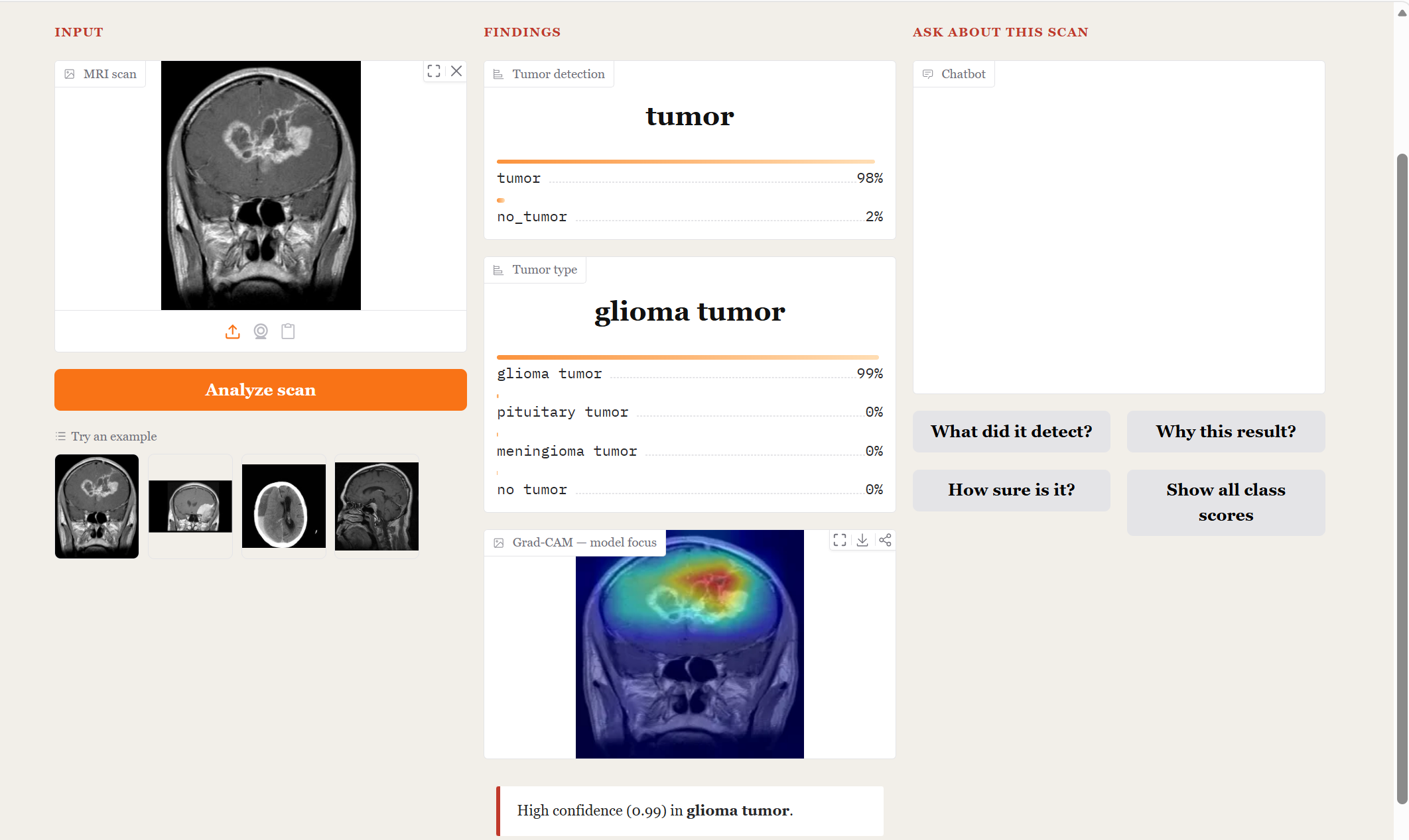
An interactive interface (Gradio) runs both models on an uploaded scan, shows a Grad-CAM overlay of where the model looked, and answers preset questions about the result — all grounded in the model's own output.
The interface:
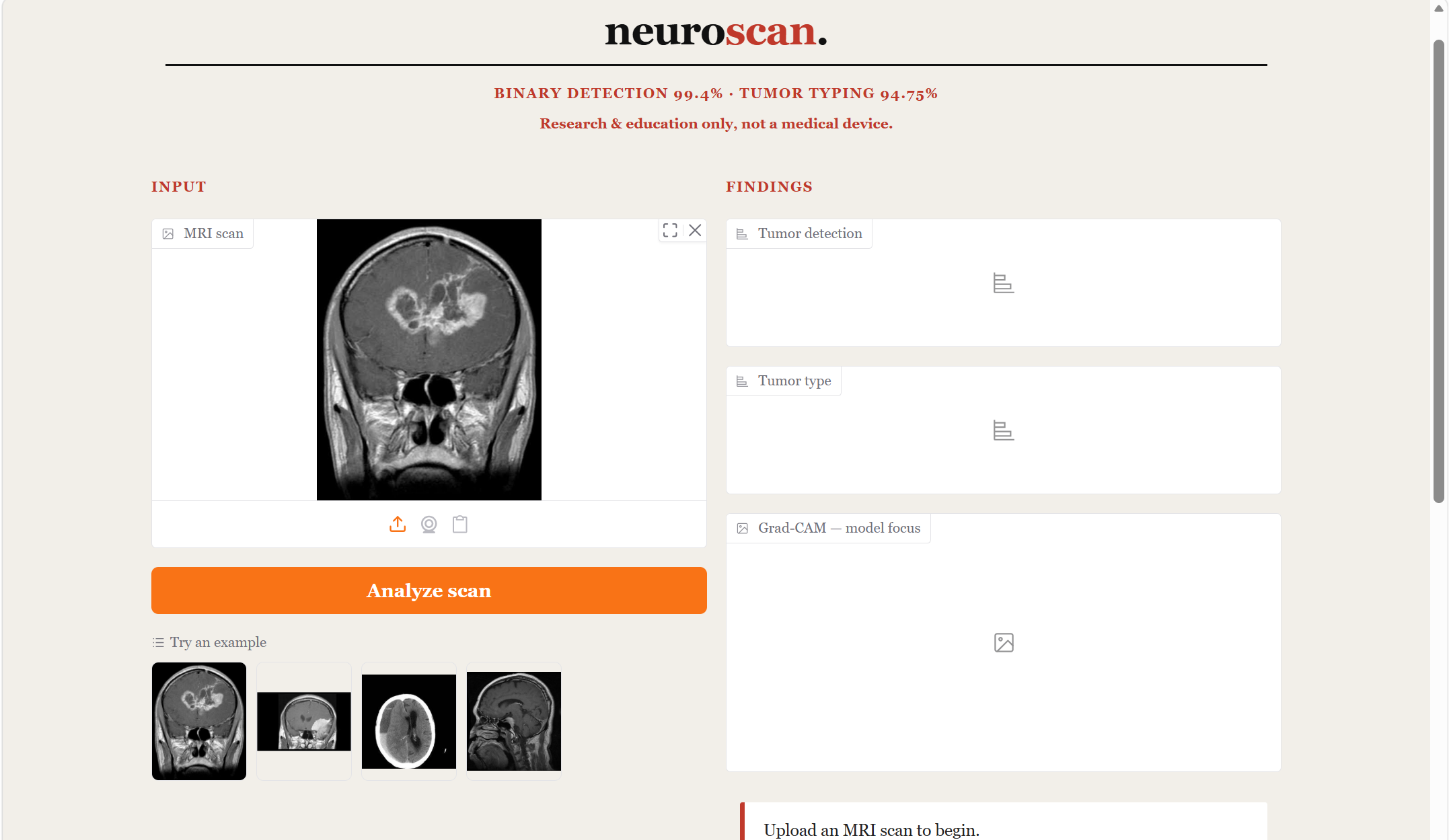
Glioma case — detection, 4-class typing, Grad-CAM, and the chat explaining the result:
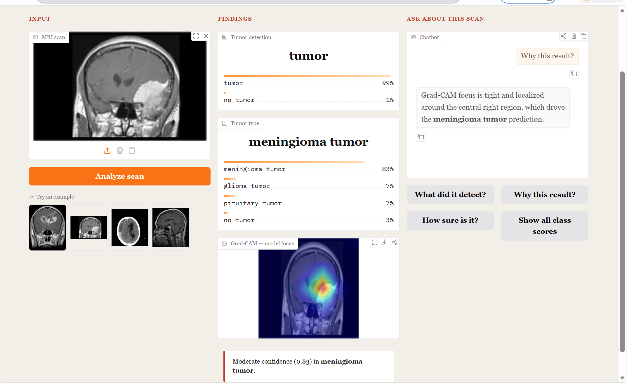
Meningioma case — the harder class, correctly typed with the chat explaining the focus region:
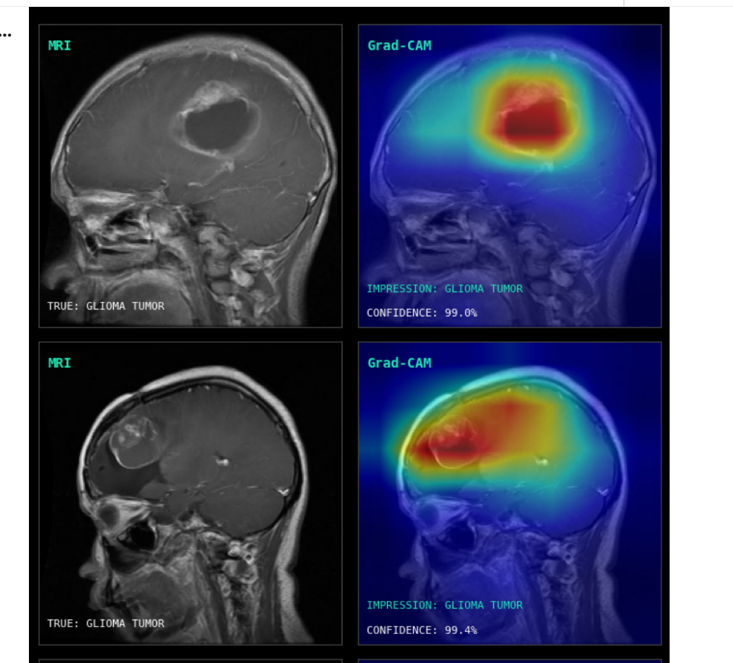
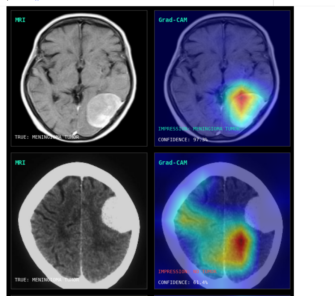
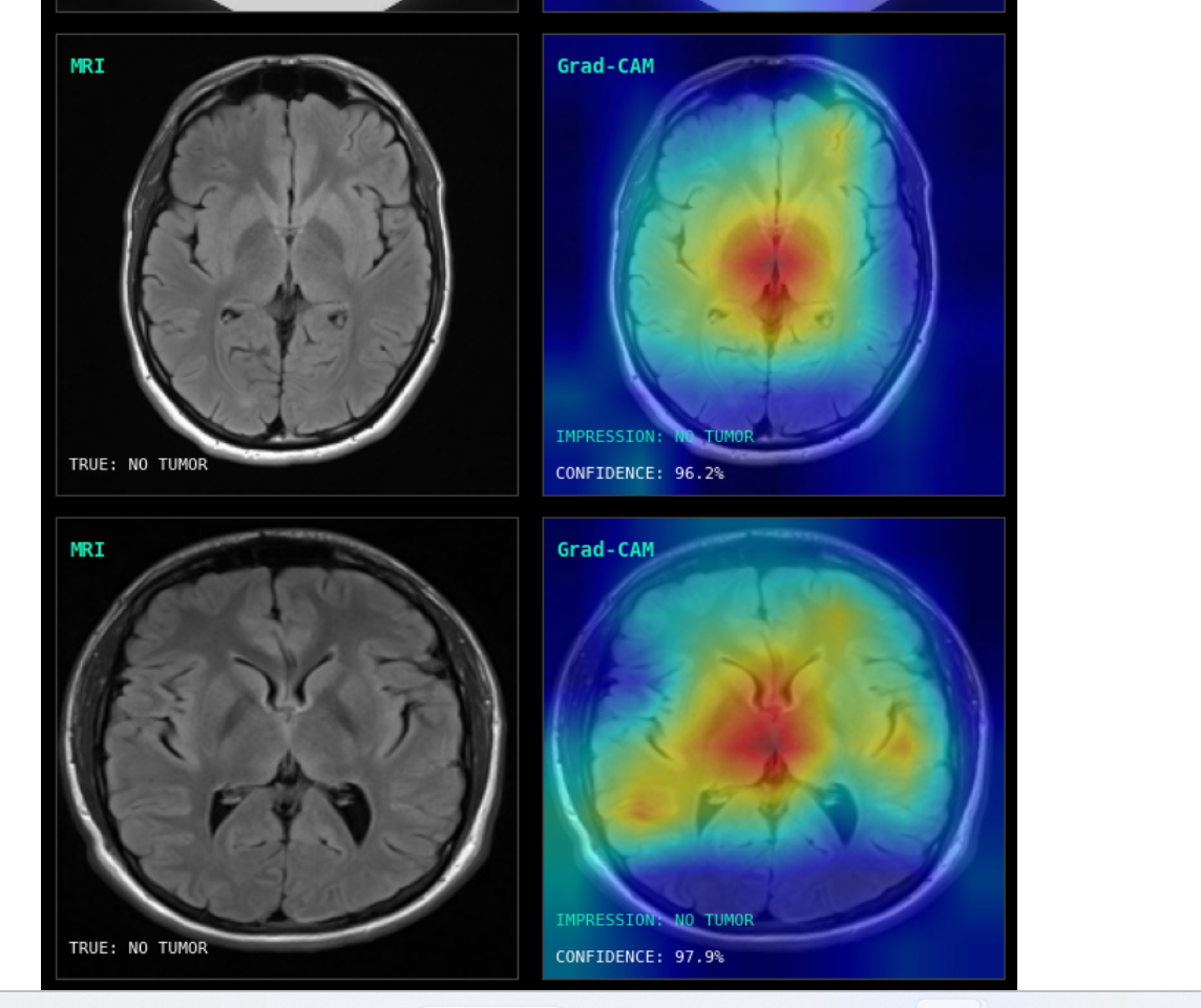
The clinical-viewer style pairs each grayscale MRI with its Grad-CAM heat map and the model's impression + confidence:
Results
Both models are EfficientNetB0, evaluated on clean stratified splits.
| Task | Accuracy | Macro-F1 |
|---|---|---|
| Binary (tumor vs no-tumor) | 99.4% | 0.988 |
| 4-class (tumor typing) | 94.75% ± 0.40% | 0.950 ± 0.004 |
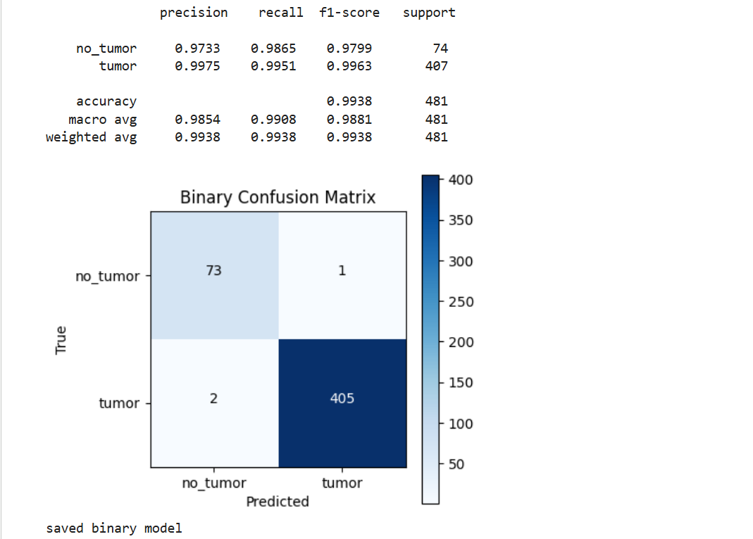
The 4-class result is averaged over 3 random seeds. On the binary task, no_tumor recall is 0.987 — of 74 healthy scans only 1 was missed — which is the key safety-relevant metric.
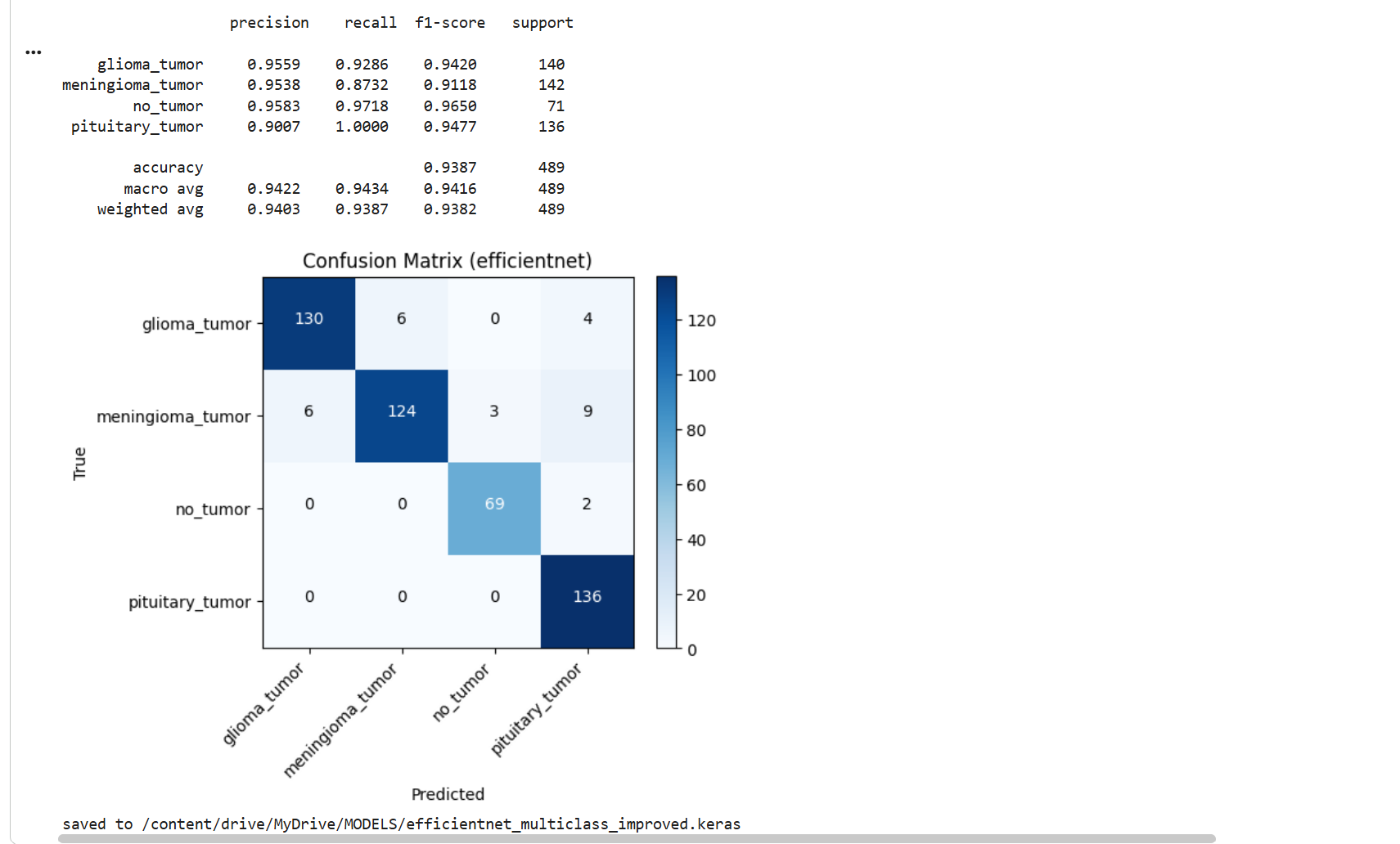
Multiclass confusion matrix:
Binary confusion matrix:
Per-class performance (representative run):
| Class | Precision | Recall | F1 |
|---|---|---|---|
| glioma | 0.96 | 0.93 | 0.94 |
| meningioma | 0.95 | 0.87 | 0.91 |
| no_tumor | 0.96 | 0.97 | 0.97 |
| pituitary | 0.90 | 1.00 | 0.95 |
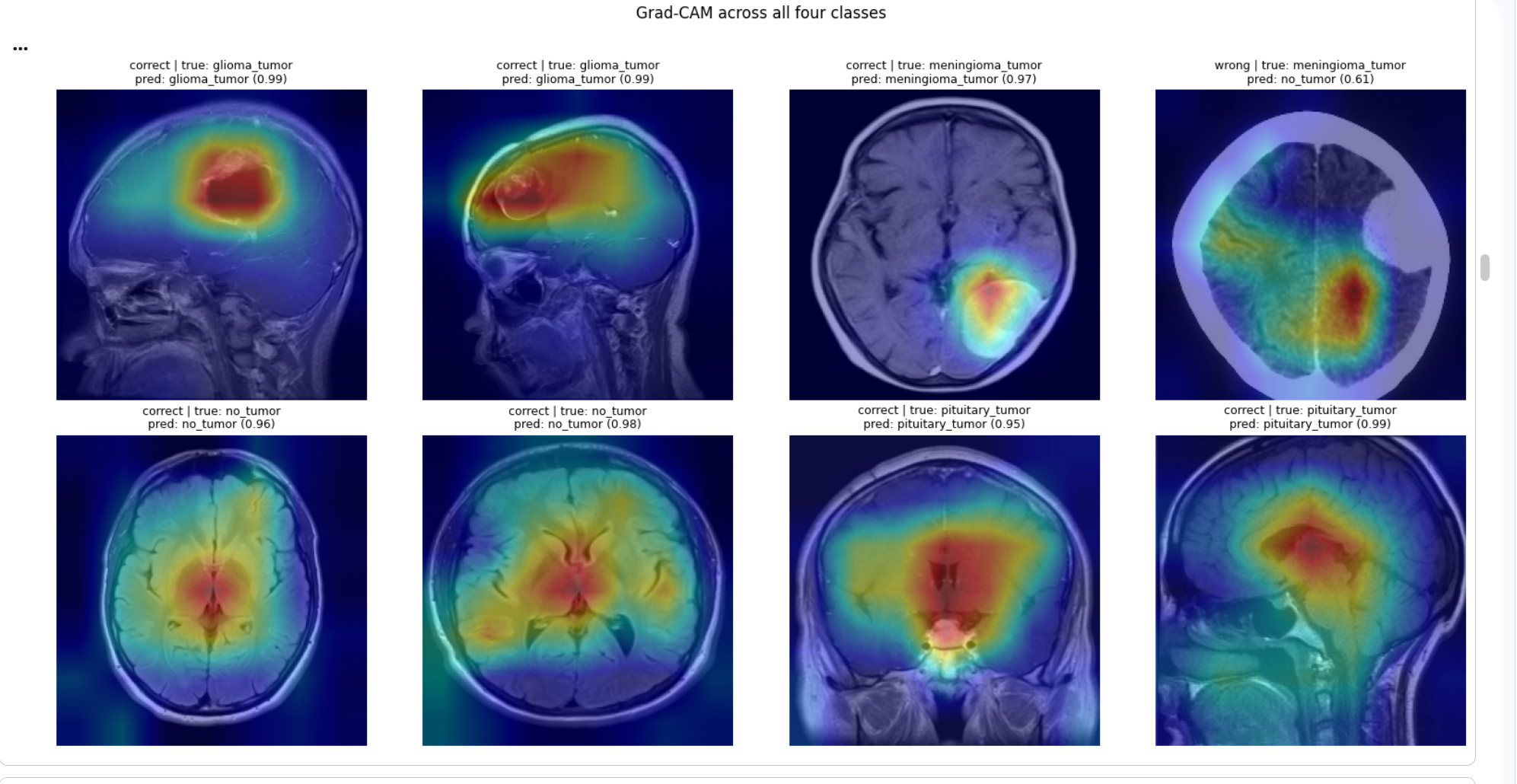
Meningioma is the hardest class, occasionally confused with glioma — consistent with their visual similarity on MRI. Grad-CAM makes this visible: on the few missed cases the heatmap is diffuse with no focal lesion, whereas correct predictions localize tightly on the tumor.
Method
- Backbone: EfficientNetB0 (ImageNet weights), ResNet50 compared as baseline.
- Input: 224×224 RGB, normalized with the backbone's own
preprocess_input. - Augmentation: horizontal flip, small rotation, zoom, and contrast, applied before normalization.
- Two-stage training: (1) train the classifier head with the backbone frozen; (2) unfreeze the upper layers and fine-tune at a low learning rate (3e-5) with cosine-style scheduling, early stopping, and LR reduction on plateau.
- Class imbalance: square-root-damped class weights (full inverse-frequency weights over-corrected and hurt minority classes).
- Loss: categorical cross-entropy with label smoothing (0.05).
- Evaluation: stratified 70/15/15 split pooled from the original folders, so train/val/test share the same distribution. Reported over 3 seeds.
Repository structure
code/
  FINAL CODE FOR BEST MODEL(MULTICLASSIFIER)/   # main 4-class notebook
  FINAL CODE FOR BEST MODEL(BINARY)/            # main binary notebook
  UI code/                                      # Gradio dual-model interface
  DATAIL CODE For Comparison/                   # baseline & comparison runs
Dataset
Brain Tumor MRI dataset with four categories: glioma, meningioma, pituitary, and no-tumor. Images are organized into class subfolders. In this project the original train/val/test folders are pooled and re-split with stratification to avoid distribution mismatch between the provided splits.
Expected layout after unzip:
DATA/
  Training(70%)/<class>/*.jpg
  Validation(20%)/<class>/*.jpg
  Testing(10%)/<class>/*.jpg
Running (Google Colab)
- Mount Drive and unzip the dataset:
from google.colab import drive drive.mount('/content/drive') !unzip -o -q "/content/drive/MyDrive/DATA.zip" -d "/content/" - Set a GPU runtime (Runtime → Change runtime type → T4 GPU).
- Open the multiclass notebook and run the cells in order: re-split → train → evaluate → Grad-CAM.
Interpretability
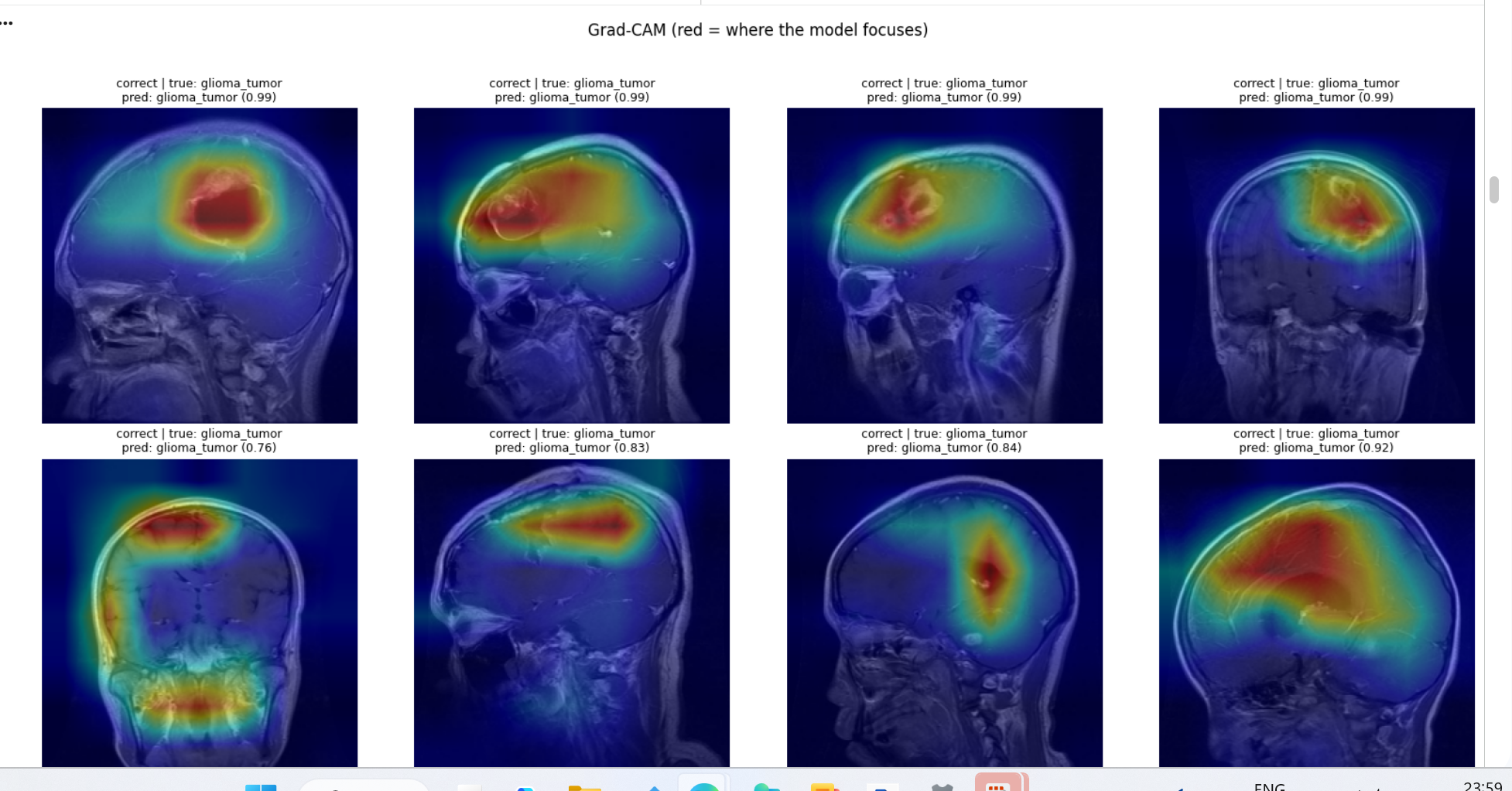
Grad-CAM is generated from the backbone's last convolutional layer and overlaid on the input scan (a grayscale clinical-viewer style is also provided). This confirms the model localizes to tumor tissue rather than skull or scanner artifacts, and explains where and why misclassifications occur.
Multiple glioma cases, showing consistent focus on the tumor region:
Limitations
- Trained and evaluated on a single public dataset; cross-dataset generalization is not yet tested.
- Not calibrated or clinically validated. Research use only.